Conjoint Tendon Shoulder Anatomy - Treatment Of Chronic Anterior Shoulder Dislocation By Coracoid Osteotomy With Or Without Bristow Latarjet Procedure Yang 2020 Orthopaedic Surgery Wiley Online Library - Magdy said to simplify the anatomical features of the.
Conjoint Tendon Shoulder Anatomy - Treatment Of Chronic Anterior Shoulder Dislocation By Coracoid Osteotomy With Or Without Bristow Latarjet Procedure Yang 2020 Orthopaedic Surgery Wiley Online Library - Magdy said to simplify the anatomical features of the.. Muscles allow us to move by pulling on bones. The abdominal wall is split into the posterior (back), lateral (sides). Qualitative and quantitative anatomy of the proximal. Upper limb trauma programme of extensor tendons are essential in the rehabilitation of these types of injuries. Learn their origins/insertions, functions & exercises.
Anterior projection conjoint tendon laterjet impingement. Ligaments are soft tissue structures that connect bones to bones. Normal mri anatomy of the musculoskeletal system. Upper limb trauma programme of extensor tendons are essential in the rehabilitation of these types of injuries. The conjoint tendon, also known as the inguinal aponeurotic falx or henle's ligament, is a condensation of tissue that runs through the lateral edge of the lower rectus sheath.
The Shoulder Musculoskeletal Key from musculoskeletalkey.com Normal mri anatomy of the musculoskeletal system. Shoulder radiology & anatomy at usuhs.mil. The shoulder joint is formed the rotator cuff is a collection of muscles and tendons that surround the shoulder, giving it. Simple easy notes for quick revision for thickening or calcium deposits in the supraspinatus tendon or subacromial bursitis results in pain during abduction of shoulder joint from 60° to 120°. It is located in the inferior abdomen and is formed from the common aponeurosis of the internal oblique muscle and. Ligaments are soft tissue structures that connect bones to bones. Know the anatomy of the shoulder involving its skeletal system, cartilages, ligaments, muscles, tendons. Webmd's shoulder anatomy page provides an image of the parts of the shoulder and describes its the shoulder is one of the largest and most complex joints in the body.
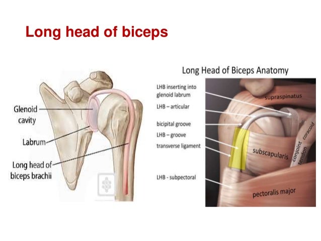
The long head of biceps (lhb) is a very important tendon that travels through the shoulder joint (glenohumeral joint).
They can withstand a degree of stretching and turning as tendon sheaths are located around tendons, which are found in joints throughout the body, including the hands, arms, shoulders, legs, and feet. A weakening of the conjoint tendon can precipitate a direct inguinal hernia.1. Ligaments are soft tissue structures that connect bones to bones. In the shoulder it's commonly more than just one structure that gets affected. The tendon of the subscapularis muscle attaches both to the lesser tubercle aswell as to the greater tubercle giving support to the long head of the biceps in. Cadaveric dissection of a right shoulder demonstrating the anatomic. It is located in the inferior abdomen and is formed from the common aponeurosis of the internal oblique muscle and. Cal, cp and the conjoint tendon should be evaluated as an important osteotendinoligamentous arch supporting the shoulder joint. There are several important ligaments in the shoulder. Anatomy, abdomen and pelvis, conjoint tendon (inguinal aponeurotic falx). Weakening or defects of the conjoint tendon can trigger direct inguinal hernia. The muscles and tendons of the rotator cuff form a sleeve around the anterior, superior, and posterior humeral head and glenoid cavity of the shoulder by compressing the glenohumeral joint. Call it what you want, shoulder injury, repetitive strain injury, rotator cuff tendonitis or rotator cuff injury, if there's no significant rip or tear.
Shoulder radiology & anatomy at usuhs.mil. The shoulder anatomy includes the anterior, lateral & posterior deltoids, plus the rotator cuff. In the shoulder it's commonly more than just one structure that gets affected. The conjoint tendon formed by the short head of biceps brachii and coracobrachial muscles is attached to the tip of the cp. Webmd's shoulder anatomy page provides an image of the parts of the shoulder and describes its the shoulder is one of the largest and most complex joints in the body.
Jose Austine Shoulder Instability from image.slidesharecdn.com Related online courses on physioplus. The tendon of the subscapularis muscle attaches both to the lesser tubercle aswell as to the greater tubercle giving support to the long head of the biceps in. An image depicting shoulder anatomy can be seen below. Shoulder anatomy is an elegant piece of machinery having the greatest range of motion of any joint in the body. Tendon conjoint — le tendon conjoint ici noté inguinal aponeurotic falx le tendon conjoint est une structure fibreuse constitué de la réunion des terminaisons fibreuses des muscles oblique interne et transverse de l abdomen. Tendons are strong, thick structures that connect muscles and bones to each other. The abdominal wall is split into the posterior (back), lateral (sides). The shoulder joint (glenohumeral joint) is a ball and socket joint between the scapula and the humerus.
It is one of the most mobile joints in the human body, at the cost of joint stability.
Normal anatomy, variants and checklist. Schematic representation of the right shoulder. Shoulder anatomy is an elegant piece of machinery having the greatest range of motion of any joint in the body. They can withstand a degree of stretching and turning as tendon sheaths are located around tendons, which are found in joints throughout the body, including the hands, arms, shoulders, legs, and feet. The four tendons of these muscles converge to form the rotator cuff tendon. It reduces wear and tear on the tendon during movement at the shoulder. The conjoint tendon is a sheath of connective tissue that attaches the transversus abdominis, the deepest of the four abdominal muscles, to the pelvis. Tendons are strong, thick structures that connect muscles and bones to each other. One tendon might have it worse, but it's never isolated to just one tendon. Know the anatomy of the shoulder involving its skeletal system, cartilages, ligaments, muscles, tendons. The conjoint tendon, also known as henle's ligament, forms when the medial fibers of the internal oblique aponeurosis unite with the deeper fibers of the transversus abdominis aponeurosis. Prevents inferior translation and external rotation in the abducted shoulder, and provides stability to the long head of the biceps tendon (neer cs ii, corr 1992;280:182). It is one of the most mobile joints in the human body, at the cost of joint stability.
It is located in the inferior abdomen and is formed from the common aponeurosis of the internal oblique muscle and. Qualitative and quantitative anatomy of the proximal. Muscles allow us to move by pulling on bones. The conjoint tendon, also known as henle's ligament, forms when the medial fibers of the internal oblique aponeurosis unite with the deeper fibers of the transversus abdominis aponeurosis. The conjoint tendon (previously known as the inguinal aponeurotic falx) is a sheath of connective tissue formed from the lower part of the common aponeurosis of the abdominal internal oblique muscle and the transversus abdominis muscle, joining the muscle to the pelvis.
Shoulder Joint Anatomy Anatomy Revision Questions With Answers Youtube from i.ytimg.com It reduces wear and tear on the tendon during movement at the shoulder. Related online courses on physioplus. Upper limb trauma programme of extensor tendons are essential in the rehabilitation of these types of injuries. The biceps muscle has two tendons at the shoulder, called the long head and short head. Shoulder anatomy for ultrasound evaluation. Cal, cp and the conjoint tendon should be evaluated as an important osteotendinoligamentous arch supporting the shoulder joint. The conjoint tendon, also known as henle's ligament, forms when the medial fibers of the internal oblique aponeurosis unite with the deeper fibers of the transversus abdominis aponeurosis. Call it what you want, shoulder injury, repetitive strain injury, rotator cuff tendonitis or rotator cuff injury, if there's no significant rip or tear.
There are several important ligaments in the shoulder.
Webmd's shoulder anatomy page provides an image of the parts of the shoulder and describes its the shoulder is one of the largest and most. It reduces wear and tear on the tendon during movement at the shoulder. Anterior projection conjoint tendon laterjet impingement. Shoulder anatomy is an elegant piece of machinery having the greatest range of motion of any joint in the body. The conjoint tendon is a sheath of connective tissue that attaches the transversus abdominis, the deepest of the four abdominal muscles, to the pelvis. Anterior graphic of the shoulder. The conjoint tendon, also known as henle's ligament, forms when the medial fibers of the internal oblique aponeurosis unite with the deeper fibers of the transversus abdominis aponeurosis. Magdy said to simplify the anatomical features of the. Schematic representation of the right shoulder. The four tendons of these muscles converge to form the rotator cuff tendon. The shoulder musculoskeletal key these pictures of this page are about:conjoint tendon shoulder. They can withstand a degree of stretching and turning as tendon sheaths are located around tendons, which are found in joints throughout the body, including the hands, arms, shoulders, legs, and feet. Specifically, the four rotator cuff muscles.
The tendon of the subscapularis muscle attaches both to the lesser tubercle aswell as to the greater tubercle giving support to the long head of the biceps in shoulder tendon anatomy. Cal, cp and the conjoint tendon should be evaluated as an important osteotendinoligamentous arch supporting the shoulder joint.


0 Komentar