Glaucoma Optic Nerve / Essentially, it's what allows the brain to receive messages from the eyes.. Nerves are delicate, and they can't stand the pressure. In glaucoma, eye pressure plays a role in damaging the delicate nerve fibers of the optic nerve. Much like alzheimer's disease is a neurodegenerative disease of the brain, glaucoma is considered a neurodegenerative disorder of the optic nerve. Traditionally, glaucoma has been viewed as a primary optic nerve disease in which the optic nerve is damaged as a result of high intraocular pressure. This optic nerve damage can eventually result in severe vision loss.
The symptoms can start so slowly that you may not notice them. Find out if we can help. We compare our findings to those for the fellow, normal nerve. Glaucoma is an umbrella term, which covers a group of diseases with a characteristic pattern of optic nerve damage. Glaucoma is one of the leading causes of blindness for people over the age of 60.
Glaucoma Glaucoma Cataract Care London from glaucoma-cataractcarelondon.co.uk If the optic nerve has abnormalities, it's likely a sign of glaucoma. How does the optic nerve work? It gets worse over time. Glaucoma is a group of eye conditions that damage the optic nerve, the health of which is vital for good vision. A layer of cells on the retina, called retinal ganglion cells, is one end of this cable. The optic nerve is composed of approximately 1.5 million axons that connect the retina to the visual targets in the brain. Much like alzheimer's disease is a neurodegenerative disease of the brain, glaucoma is considered a neurodegenerative disorder of the optic nerve. Glaucoma is one of the leading causes of blindness for people over the age of 60.
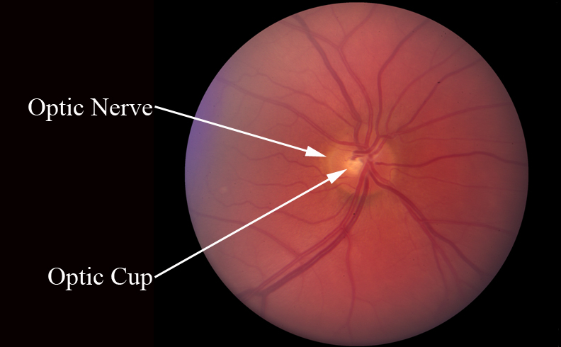
The glaucoma research foundation says glaucoma is a common cause of optic cupping.
The optic nerve is vulnerable to getting damaged by pressure in the eyes, called ocular hypertension. There are many reasons for this, including the fact that retinal ganglion cells cannot repair or regenerate. Traditionally, glaucoma has been viewed as a primary optic nerve disease in which the optic nerve is damaged as a result of high intraocular pressure. The glaucoma research foundation says glaucoma is a common cause of optic cupping. There are many types of glaucoma classified by clinical exams. Axons can be thought of as long cables or extensions of the retinal ganglion cells, which are the cells that are damaged in glaucoma. This damage is often caused by an abnormally high pressure in your eye. 700+ patients with optic nerve damage have regained lost vision. Glaucoma can cause the cup to enlarge (actually little nerve fibers are being wiped out along the rim of the optic nerve in glaucoma). This makes sense, since many patients with apparent glaucoma present in clinic with high pressure—and in most of those cases, if you lower the iop the patient stops progressing. If the optic nerve has abnormalities, it's likely a sign of glaucoma. Glaucoma results in progressive damage to the optic nerve due to high pressure in the eye (intraocular pressure). Find out if we can help.
Unfortunately, like other central nervous system regions such as the spinal cord, the regenerative capacity of the optic nerve is limited. Through periodic photographs of the optic nerve, the ratio of the cup to the disc can be monitored. The symptoms can start so slowly that you may not notice them. Glaucoma is a group of eye diseases causing optic nerve damage. Traditionally, glaucoma has been viewed as a primary optic nerve disease in which the optic nerve is damaged as a result of high intraocular pressure.
Ch 1 Glaucoma Optic Nerve Disease A Patient S Guide To Glaucoma from eyerounds.org Find out if we can help. The optic nerve is composed of approximately 1.5 million axons that connect the retina to the visual targets in the brain. The optic nerve is vulnerable to getting damaged by pressure in the eyes, called ocular hypertension. Optic nerve photographs show optic nerve damage over time. Axons can be thought of as long cables or extensions of the retinal ganglion cells, which are the cells that are damaged in glaucoma. Glaucoma is a condition that damages your eye 's optic nerve. What is the optic nerve? (image adapted from the internet) unfortunately, any vision lost from the optic nerve damage cannot be recovered.
In order to evaluate the optic nerve carefully, a clear stereoscopic view with a 78.00 or 60.00 d lens is necessary.
It can occur at any age but is more common in older adults. Glaucoma can cause the cup to enlarge (actually little nerve fibers are being wiped out along the rim of the optic nerve in glaucoma). The optic nerve carries images from the retina, which is the specialized light sensing tissue, to the brain so we can see. The optic nerve is composed of approximately 1.5 million axons that connect the retina to the visual targets in the brain. When a significant number of nerve fibers are damaged, blind. Glaucoma is an optic neuropathy leading to changes in the intrapaillary and parapaillary regions of the optic disk. Glaucoma typically causes the cup to get bigger in a vertical oval type pattern. Understanding glaucoma we rely on the optic nerve to translate messages to the brain. Glaucoma is a group of eye conditions that damage the optic nerve, the health of which is vital for good vision. This makes sense, since many patients with apparent glaucoma present in clinic with high pressure—and in most of those cases, if you lower the iop the patient stops progressing. Glaucoma is actually a group of diseases, but the common feature among all types of glaucoma is optic nerve degeneration. We've helped 700+ patients with glaucoma restore their vision. A layer of cells on the retina, called retinal ganglion cells, is one end of this cable.
When you have glaucoma, your eye is filled with an unusual amount of fluid, and it puts immense pressure on all the tissues in your eye. Optic nerve photographs show optic nerve damage over time. Optic nerve assessment contributes to the clinician's ability to detect glaucoma. Despite technological advances, clinical identification of optic nerve head characteristics remains the first step in diagnosis. There are many types of glaucoma classified by clinical exams.
Strategies For Improving Early Detection Of Glaucoma The Combined Str Opth from www.dovepress.com Crucial details include the color and contour of the neuroretinal rim and the vascular contour (eg, barring of a retinal vessel due to lost tissue in the rim or. How does the optic nerve work? If not diagnosed early enough, glaucoma can. There are many types of glaucoma classified by clinical exams. In glaucoma, eye pressure plays a role in damaging the delicate nerve fibers of the optic nerve. The optic nerve is the bundle of nerve fibers at the back of the eye that carry visual messages from the retina to the brain. In glaucoma, the axons of these optic nerve cells degenerate, and eventually lead to cell death. The only way to find out if you have glaucoma is to get a comprehensive dilated eye exam.
Glaucoma typically causes the cup to get bigger in a vertical oval type pattern.
Glaucoma and optic nerve repair cell tissue res. How does the optic nerve work? Axons can be thought of as long cables or extensions of the retinal ganglion cells, which are the cells that are damaged in glaucoma. A layer of cells on the retina, called retinal ganglion cells, is one end of this cable. Optic nerve assessment contributes to the clinician's ability to detect glaucoma. Careful examination of the disk parameters including si … The glaucoma research foundation says glaucoma is a common cause of optic cupping. The optic nerve is a cable of nerve fibers that carry electrical impulses from the retina to the brain. In glaucoma, eye pressure plays a role in damaging the delicate nerve fibers of the optic nerve. In order to evaluate the optic nerve carefully, a clear stereoscopic view with a 78.00 or 60.00 d lens is necessary. Glaucoma is one of the leading causes of blindness for people over the age of 60. Find out if we can help. Your eye doctor may use one of these optic nerve computer imaging techniques as part of your glaucoma examination.
Glaucoma is a group of eye diseases which result in damage to the optic nerve (or retina) and cause vision loss glaucoma. This makes sense, since many patients with apparent glaucoma present in clinic with high pressure—and in most of those cases, if you lower the iop the patient stops progressing.


0 Komentar